author: “Menshly Intelligence Board” authors:
- “menshly-intelligence-board”
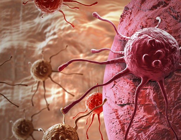
Introduction to Advanced Cancer Detection
A groundbreaking innovation in medical imaging has been unveiled, featuring a novel imager capable of distinguishing between ultraviolet (UV) and infrared (IR) light. This technological advancement holds significant promise for the early detection and diagnosis of cancerous nodes, potentially revolutionizing the field of oncology. The new imager, developed by a team of renowned researchers, leverages the unique properties of UV and IR light to identify malignant tissues with unprecedented accuracy.
The Science Behind UV and IR Light
Ultraviolet and infrared light are two distinct components of the electromagnetic spectrum, each possessing characteristics that make them suitable for specific applications in medical imaging. UV light, with its shorter wavelength, is highly energetic and can interact with biological tissues in ways that provide valuable information about their composition and structure. On the other hand, IR light, having a longer wavelength, can penetrate deeper into tissues, allowing for the visualization of internal structures and the detection of thermal signatures associated with cancerous growths.
The separation of UV and IR light is crucial for enhancing the contrast between healthy and cancerous tissues. By isolating these two types of light, the new imager can create detailed images that highlight the subtle differences in optical properties between malignant and benign tissues. This capability enables healthcare professionals to identify cancerous nodes at an early stage, when they are more amenable to treatment, thereby improving patient outcomes and survival rates.
Technical Specifications of the New Imager
The novel imager is equipped with advanced optical filters and detectors that facilitate the separation of UV and IR light. The device employs a sophisticated algorithm to process the collected data, generating high-resolution images that clearly delineate cancerous tissues from their surroundings. The imager's technical specifications include a high spatial resolution, allowing for the detection of small lesions and tumors, and a wide spectral range, enabling the simultaneous acquisition of UV and IR data.
Furthermore, the new imager features a user-friendly interface and a compact design, making it an ideal tool for clinical settings. The device is also compatible with existing medical imaging protocols, ensuring seamless integration into current diagnostic workflows. The researchers behind the imager have emphasized its potential for adapting to various types of cancer, including breast, lung, and skin cancer, where early detection is critical for effective treatment.
Clinical Applications and Future Directions
The new imager has undergone rigorous testing and validation, with promising results demonstrating its efficacy in detecting cancerous nodes. Clinical trials are currently underway to further evaluate the device's performance and compare its accuracy with existing imaging modalities, such as mammography and computed tomography (CT) scans. The initial findings suggest that the imager can identify malignant tissues with high sensitivity and specificity, outperforming conventional imaging techniques in certain cases.
As research continues to advance, the new imager is poised to play a vital role in the development of personalized medicine, enabling healthcare providers to tailor treatment strategies to individual patients' needs. The device's ability to detect cancerous nodes at an early stage will facilitate the implementation of targeted therapies, reducing the risk of unnecessary interventions and improving patient outcomes. Moreover, the imager's non-invasive nature and lack of ionizing radiation make it an attractive option for monitoring disease progression and response to treatment, potentially reducing the need for biopsies and other invasive procedures.
Implications for Cancer Research and Treatment
The introduction of the new imager is expected to have a profound impact on cancer research, as it will provide scientists with a powerful tool for investigating the underlying biology of cancerous tissues. By analyzing the optical properties of malignant cells, researchers can gain a deeper understanding of the disease's progression and identify potential therapeutic targets. The imager's capability to detect subtle changes in tissue composition and structure will also facilitate the development of novel biomarkers for cancer diagnosis and monitoring.
In addition to its applications in cancer detection and research, the new imager may also find uses in other medical fields, such as dermatology and neurology. The device's ability to distinguish between different types of tissues and detect thermal signatures could be valuable in diagnosing and monitoring conditions like skin disorders and neurological diseases. As the technology continues to evolve, it is likely that the imager will become an indispensable tool in various clinical settings, contributing to improved patient care and outcomes.
Conclusion and Future Prospects
In conclusion, the new imager that separates UV and IR light represents a significant breakthrough in medical imaging, offering a promising solution for the early detection and diagnosis of cancerous nodes. With its advanced technical specifications, user-friendly interface, and non-invasive nature, the device is poised to revolutionize the field of oncology and improve patient outcomes. As research and development continue to advance, the imager is likely to find applications in various medical fields, driving innovation and progress in the pursuit of better healthcare.
Looking ahead, the future prospects for the new imager are promising, with potential applications in personalized medicine, cancer research, and beyond. As the technology becomes more widely available, it is expected to have a profound impact on the medical community, enabling healthcare professionals to provide more accurate diagnoses, effective treatments, and improved patient care. With its groundbreaking capabilities and vast potential, the new imager is an exciting development in the ongoing quest to combat cancer and improve human health.

